Zubehör mit
Mikroskopmodul (MMA)
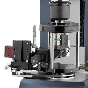
Ermöglicht umfassende Strömungsvisualisierung – einschließlich Gegenlauf – mit gleichzeitigen rheologischen Messungen auf einem Discovery Hybrid Rheometer.
Modular Microscope Accessory (MMA)
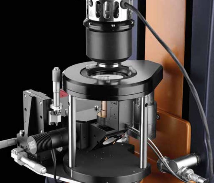
Das Zubehör mit Mikroskopmodul (MMA) ermöglicht eine umfassende Strömungsvisualisierung – einschließlich Gegenlauf – mit gleichzeitigen rheologischen Messungen auf einem Discovery Hybrid Rheometer. Eine hochauflösende Kamera erfasst Bilder mit bis zu 90 FPS über standardmäßige Mikroskopobjektive mit einer bis zu 100-fachen Vergrößerung. Die Beleuchtung durch eine blaue LED kann zur selektiven Beleuchtung oder für die Fluoreszenzmikroskopie mit einem Kreuzpolarisator oder einem dichroitischen Teiler gekoppelt werden.
 Technologie
Das MMA wird direkt am Discovery Hybrid Rheometer angebracht und erfordert keine zusätzlichen Stative, Hebevorrichtungen oder sonstigen Halterungen. Dadurch ist das System einfach zu installieren und effektiv von externer Vibration und anderen Umwelteinflüssen isoliert, die die Bildqualität beeinträchtigen können. Dank des hochpräzisen x-y-z-Mikrometerpositionierungssystems lässt sich das Sichtfeld des Mikroskops auf eine beliebige Stelle der Probe richten. Dies ermöglicht eine Untersuchung der Strömungshomogenität an einer beliebigen Position von der Rotationsachse bis zum Rand der Probe. Die präzise Tiefenprofilierung wird durch ein optionales Piezo-Scansystem gewährleistet. Mithilfe dieses Präzisionsmechanismus lässt sich die die Tiefe der Fokusebene über einen Bereich von 100 µm in softwaregesteuerten Schritten von nur 0,1 mm zur quantitativen Tiefenprofilierung einstellen. Das MMA ist kompatibel mit der oberen Heizplatte UHP zur Temperaturregelung zwischen -20 °C bis 100 °C.
Gegenlauf: Mikroskopie mit Stagnationsebene
Bei der Visualisierung fließender Materialien bei hohen Scherraten bewegen sich relevante Merkmale möglicherweise schnell durch das Sichtfeld, sodass zum Beobachten der durch die Scherung verursachten Veränderungen an der Probe nur wenig Zeit bleibt. Mit der optionalen Gegenlaufphase für das MMA wird die untere Glasplatte in konstanter Geschwindigkeit gegenläufig zur oberen Platte gedreht. Dadurch entsteht eine Stagnationsebene mit Nullgeschwindigkeit. Das heißt, das Fluid steht relativ zur Kamera still, sodass während des Versuchs ein konstantes Sichtfeld erhalten bleibt. Die Position dieser Nullgeschwindigkeitsfläche innerhalb des Spalts lässt sich einstellen, indem das Verhältnis der Geschwindigkeiten der oberen und unteren Platte variiert wird, ohne die effektive Schergeschwindigkeit an der Probe zu ändern. Bei diesem Gegenlaufsystem handelt es sich um ein Smart Swap™-Zubehör, das jederzeit hinzugefügt werden kann.
|
Kreuzpolarisator |
Enthalten |
|
Dichroitischer Fluoreszenz-Teiler |
Optional |
|
Gegenlauf |
Optionales Smart Swap™ System |
|
Piezo-Scanmechanismus |
Optional, Verfahrweg von 100 µm |
|
Video- und Bildaufnahme |
Software-gesteuert, Datendatei integriert |
|
Sichtfeld |
320 mm x 240 mm bei 20-fach |
|
Beleuchtung |
Blaue LED |
|
Bilderfassung |
640 x 480 Pixel, 90 FPS |
|
Temperaturbereich (mit UHP) |
-20 °C bis 100 °C |
|
Geometrien |
Platten und Kegel mit Durchmessern von bis zu 40 mm |
Merkmale und Vorteile
- Smart Swap™-Technologie zur schnellen Installation
- Gegenlauf zur Stagnationsebenenabbildung auf einem beliebigen Discovery Hybrid Rheometer
- Hohe Auflösung und Bildrate
- Effektive Temperaturregelung durch die obere Heizplatte UHP
- Direkte Messung der Probentemperatur mit der aktiven Temperaturregelung ATC
- Sicht auf jede Position innerhalb der Messfläche, z. B. Mittelpunkt, Rand oder Mittenradius
- Optionale Kreuzpolarisierung, Fluoreszenz und präzise Tiefenprofilierung
- Breite Auswahl an gewerblich erhältlichen Objektiven
- Beschreibung
-
Modular Microscope Accessory (MMA)
Das Zubehör mit Mikroskopmodul (MMA) ermöglicht eine umfassende Strömungsvisualisierung – einschließlich Gegenlauf – mit gleichzeitigen rheologischen Messungen auf einem Discovery Hybrid Rheometer. Eine hochauflösende Kamera erfasst Bilder mit bis zu 90 FPS über standardmäßige Mikroskopobjektive mit einer bis zu 100-fachen Vergrößerung. Die Beleuchtung durch eine blaue LED kann zur selektiven Beleuchtung oder für die Fluoreszenzmikroskopie mit einem Kreuzpolarisator oder einem dichroitischen Teiler gekoppelt werden.
- Technologie
-
 Technologie
Das MMA wird direkt am Discovery Hybrid Rheometer angebracht und erfordert keine zusätzlichen Stative, Hebevorrichtungen oder sonstigen Halterungen. Dadurch ist das System einfach zu installieren und effektiv von externer Vibration und anderen Umwelteinflüssen isoliert, die die Bildqualität beeinträchtigen können. Dank des hochpräzisen x-y-z-Mikrometerpositionierungssystems lässt sich das Sichtfeld des Mikroskops auf eine beliebige Stelle der Probe richten. Dies ermöglicht eine Untersuchung der Strömungshomogenität an einer beliebigen Position von der Rotationsachse bis zum Rand der Probe. Die präzise Tiefenprofilierung wird durch ein optionales Piezo-Scansystem gewährleistet. Mithilfe dieses Präzisionsmechanismus lässt sich die die Tiefe der Fokusebene über einen Bereich von 100 µm in softwaregesteuerten Schritten von nur 0,1 mm zur quantitativen Tiefenprofilierung einstellen. Das MMA ist kompatibel mit der oberen Heizplatte UHP zur Temperaturregelung zwischen -20 °C bis 100 °C.
Gegenlauf: Mikroskopie mit Stagnationsebene
Bei der Visualisierung fließender Materialien bei hohen Scherraten bewegen sich relevante Merkmale möglicherweise schnell durch das Sichtfeld, sodass zum Beobachten der durch die Scherung verursachten Veränderungen an der Probe nur wenig Zeit bleibt. Mit der optionalen Gegenlaufphase für das MMA wird die untere Glasplatte in konstanter Geschwindigkeit gegenläufig zur oberen Platte gedreht. Dadurch entsteht eine Stagnationsebene mit Nullgeschwindigkeit. Das heißt, das Fluid steht relativ zur Kamera still, sodass während des Versuchs ein konstantes Sichtfeld erhalten bleibt. Die Position dieser Nullgeschwindigkeitsfläche innerhalb des Spalts lässt sich einstellen, indem das Verhältnis der Geschwindigkeiten der oberen und unteren Platte variiert wird, ohne die effektive Schergeschwindigkeit an der Probe zu ändern. Bei diesem Gegenlaufsystem handelt es sich um ein Smart Swap™-Zubehör, das jederzeit hinzugefügt werden kann.
Kreuzpolarisator
Enthalten
Dichroitischer Fluoreszenz-Teiler
Optional
Gegenlauf
Optionales Smart Swap™ System
Piezo-Scanmechanismus
Optional, Verfahrweg von 100 µm
Video- und Bildaufnahme
Software-gesteuert, Datendatei integriert
Sichtfeld
320 mm x 240 mm bei 20-fach
Beleuchtung
Blaue LED
Bilderfassung
640 x 480 Pixel, 90 FPS
Temperaturbereich (mit UHP)
-20 °C bis 100 °C
Geometrien
Platten und Kegel mit Durchmessern von bis zu 40 mm
Merkmale und Vorteile
- Smart Swap™-Technologie zur schnellen Installation
- Gegenlauf zur Stagnationsebenenabbildung auf einem beliebigen Discovery Hybrid Rheometer
- Hohe Auflösung und Bildrate
- Effektive Temperaturregelung durch die obere Heizplatte UHP
- Direkte Messung der Probentemperatur mit der aktiven Temperaturregelung ATC
- Sicht auf jede Position innerhalb der Messfläche, z. B. Mittelpunkt, Rand oder Mittenradius
- Optionale Kreuzpolarisierung, Fluoreszenz und präzise Tiefenprofilierung
- Breite Auswahl an gewerblich erhältlichen Objektiven


